At Metrolab, we are eager to learn more about our customers’ applications. Therefore, we approached Luca Bartalini, a specialist in MRI and author of the book: “Artifacts and Technical Solutions in MR Diagnostic Imaging” to write an introduction on MRI artifacts induced by magnet inhomogeneity.
Introduction
The aim of this article is to give a short overview of the world of MRI artifacts, focusing on those which are induced by magnet inhomogeneity. The structure of the article is:
- What are artifacts? definition and characteristics
- MRI artifact classification
- The magnet and its characteristics
- Magnetic field inhomogeneity artifacts
- Conclusions
What are artifacts?
Artifacts are all those imaging alterations which can modify the real anatomical and/or pathological condition. Artifacts usually manifest as anatomy distortion, anatomy shifting or ghosting.
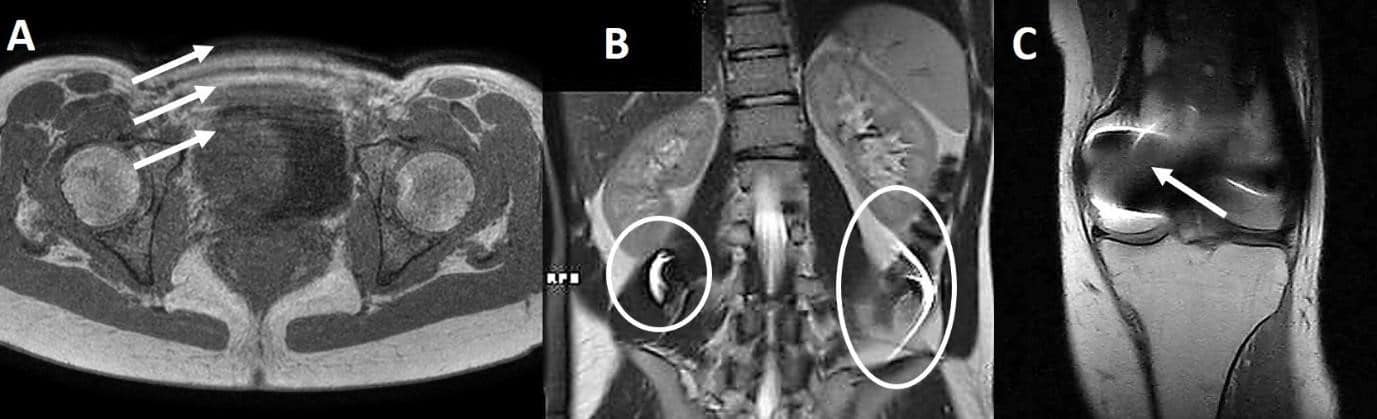
Figure 1 Examples of MRI artifact: (A) Ghosting induced by breathing, (B) Superimposed objects due to the incorrect application of Parallel Imaging and/or incorrect patient positioning, (C) Signal void and pile up artifact due to metallic implantology (Courtesy of Luca Bartalini ).
Compared to other diagnostic modalities, MRI is particularly prone to artifacts. This is largely due to the extreme complexity of MRI physical principles and to the large number of hardware components. The appearance of the artifact can change depending on its cause and on the MRI system brand: the same artifact, produced by different brands of MRI, can manifest in different ways. Such variability and complexity requires a robust knowledge on the part of the MRI technologists/radiographers in order to identify and correct for all types of artifacts, applying techniques such as parameter modification through to patient positioning. In addition, radiologists should recognize the most common artifact manifestations in order to avoid image misinterpretation, such as “magic angle”, which can change the intensity of the MRI signal without distortions, anatomy shifting or superimposed objects.

Figure 2 High signal of the supraspinatus tendon (white circle). This alteration is not a pathology but an artifactual signal alteration called the “Magic Angle”, caused by the specific value of echo time and anatomy orientation (Courtesy of Luca Bartalini).
MRI artifact classification
MRI artifacts can manifest in many ways and can be influenced by many variables. Below is a classification of MRI artifacts based on their causes:
-
Human Physiology-Related Artifacts
- Movement
- Vascular Pulsation
- Slow Flow
-
Hardware Artifacts
-
K-Space-Related Artifacts
- Aliasing
- Generic Subsampling
- Parallel Subsampling
- Non-Cartesian Advanced Fillings
-
Spin Physics-Related Artifacts
- Chemical Shift
- J-Coupling
- Spectral Tissue Saturation
-
Artifacts Related to Specific Acquisition Techniques
- Magic Angle
- Pseudolayering CA
- Dixon
- Steady State Free Precession (SSFP)
- EPI-DWI
- MR Angiography (with and without contrast media)
- Functional MRI
-
New Technology-Related Artifacts…
The magnet and its characteristics
The magnet is “the heart” of the MRI scanner. It creates the static magnetic field B0 which generates the conditions necessary to create the magnetic resonance phenomenon. The magnet has three principal characteristics:
- Intensity (TeslaThe SI unit for magnetic flux density (B). More).
- Temporal stability.
- Spatial stability or homogeneity.

Figure 3 On the left is a perfect magnet homogeneity with rectilinear magnetic lines (arrows). On the right is a remarkable inhomogeneity with the modification of field lines.
In MRI, magnet homogeneity is the most important characteristic, because it facilitates the correct and homogeneousA magnetic field with no or only a very small gradient. NMR teslameters can only be used in fields that... More absorption of the radio frequency pulse, without signal void and anatomy distortion. The process to improve magnet homogeneity is called “shimmingIn magnet production, the process of making a magnetic field more homogeneous, by placing pieces of iron (shims) in the... More”. It can be “passive”, through static implementation or “active”, using specific coils. The active method can improve the quality of the image, in particular when spectral saturation or Diffusion Weighted Imaging acquisitions are requested.
— Introduced 25 years ago, Metrolab’s NMRNuclear Magnetic Resonance. A resonance phenomenon seen when you irradiate a sample in a magnetic field with an RF field.... More Magnetic Field Cameras (MFC) have revolutionized field mappingThe process of measuring magnetic field intensity at many different points, in order to understand the structure of the field... More for MRI magnets. The maps acquired using the MFC enable the manufacturer to shim their magnets and reduce the static magnetic field B0 inhomogeneity. This process occurs during the production of the magnet as well as later during the installation of the MRI. MFC, like the MFC2046, reduce acquisition times from hours to minutes, positioning errors to fractions of a millimeter, and they render human and driftThe gradual loss of an instrument's accuracy. NMR teslameters drift because their time base drifts; this can be easily checked and... More errors negligible. —
Note by Metrolab
Magnetic field inhomogeneity artifacts
Magnet inhomogeneity can induce artifacts, based on the type of sequence, the scan parameters, receiver coil type and patient positioning. The most common are:
- Anatomy distortion
- Spectral saturation errors
- Signal to Noise Ratio (SNR) reduction
- Dixon Swap
- Other
1) Anatomy distortion is a recurrent artifact that occurs when a metallic object, such as an articular prosthesis, is inserted into the magnetic field. The distortion may also become visible in the absence of metallic implants, especially in the periphery of the maximum field of view.
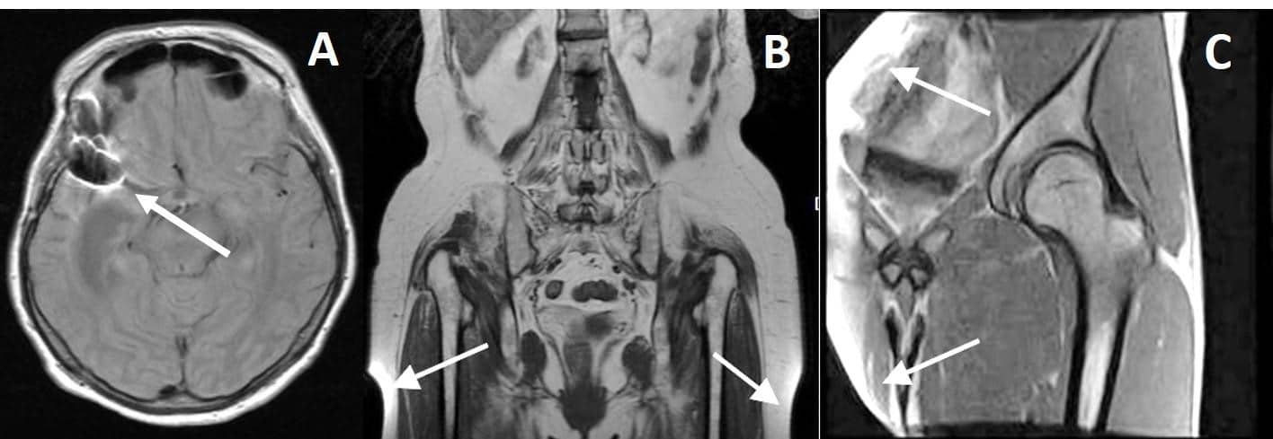
Figure 4 Magnetic field inhomogeneity: (A) Susceptibility artifact due to a metallic implant, (B) Peripheral anatomy distortion at maximum Field Of View (FOV), (C) Strong distortion with small FOV due to marked inhomogeneity (Courtesy of Luca Bartalini).
2) Different types of spectral saturations are often used clinically, with or without contrast media. They are all prone to field inhomogeneity.
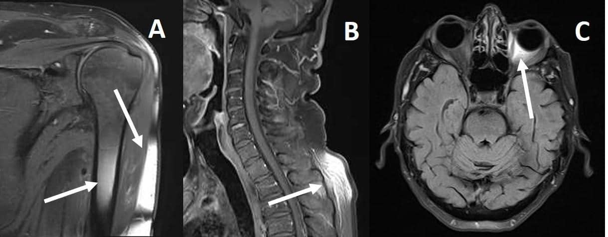
Figure 5 Fat saturation failure due to field inhomogeneity. The case “C” can be very dangerous for medical interpretation (Courtesy of Luca Bartalini and Giuseppe Walter Antonucci).
3) Diffused magnet inhomogeneity can reduce SNR. It is a non-specific signal which can be induced by many different factors. 4) The Dixon technique is commonly used to produce pictures with strong fat subtraction. It is based on a post processing algorithm which creates a fat and water series, starting from multi-echo acquisition: magnet inhomogeneity can produce a specific artifact called “Swap”.

Figure 6 The Dixon swap during the post processing due to inhomogeneity (Courtesy of Alan Gerevini).
5) The artifacts described above are the most common in clinical practice, but inhomogeneity can create other artifactual situations not recorded here.
Conclusions
MRI artifacts are very common clinical occurrences and require the radiographer/technologist to recognize and account for each artifact, in every condition. There are many technical parameters that can be changed to solve or reduce artifacts, but their treatment goes beyond the scope of this article. Artifacts due to the magnet inhomogeneity are very common and sometimes hard to recognize. The quality of the magnet and its homogeneity must be perfect in order to provide a high-level image in any condition. Of course, this must be complemented by a high standardA standard is the internationally agreed-upon physical representation of a unit. For example, a caesium clock is the standard for... More of technical and medical competence.